About this deal
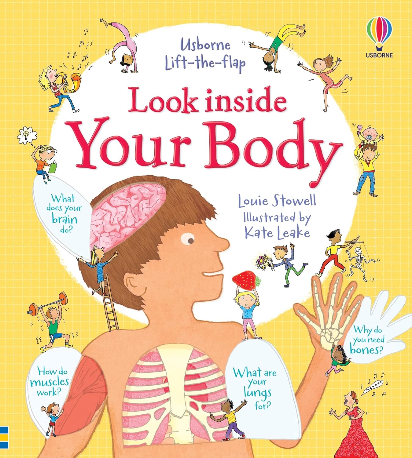
This book is so great. It is so informative. My son loves to look and see what happens when you eat food (let’s be real – he loves to see the poop)! It has over 100 flaps (including flaps in flaps)! It shows how your muscles work and how your brain processes what you see. I highly recommend this book.” Other Usborne Books & More titles in the Series include: Ultrasound scanners were not commonly used in hospitals until the 1970s. By the 1980s the technology had advanced enough to produce moving images in shades of grey, followed by 3D imaging not long after. Today ultrasound is widely used in surgical procedures and the field of gynaecology. Capsule endoscopy is a procedure that uses a tiny wireless camera to take pictures of the digestive tract. It helps practitioners see inside the small intestine—an area that isn't easily reached with a traditional endoscopy procedure. X-rays were the first technology that made it possible to see inside the body without having to open it up. They were discovered by German physicist Wilhelm Roentgen (1845–1923) at the end of the 1800s and had an immediate impact on anatomical study and diagnostics.
Look Inside: Your Body - Louie Stowell - Google Books Look Inside: Your Body - Louie Stowell - Google Books
X-rays are a high-energy, invisible, form of electromagnetic radiation. Like visible light, they are reflected by some objects and absorbed to varying degrees by others. Peek under all the flaps in these colorful and engaging books–perfect for little fingers and curious minds.”– Usborne Look Inside Your Body Inside Look Inside Your Body It is written in an informative, factual but informal way which is beneficial because it is adding to the children’s vocabulary as well as understanding things that are going on with their own bodies in a fun way. Your Body Uncovered with Kate Garraway’ on BBC2 uses virtual reality to allow people to take a look inside themselves.Because MRI can construct images of soft tissue, it's especially useful for diagnosing joint abnormalities, diseases of the liver and abdominal organs, and identifying tumours and uterine conditions such as fibroids. Dr Singh says: "The 3D images of the patients’ bodies are incredible. As a doctor, it sometimes feels that language isn’t up to the job of truly explaining what is happening inside the gloriously complex human body. How amazing, then, to have a tool that allows realistic, accurate and personal images to do some of the job for you! When you’re ill, the first step towards recovery is coming to terms with your diagnosis, and I think being able to see, literally, what’s going on inside your own body gives patients the understanding to really do that. But this isn’t just for patients – the truth is, despite all my years of medical training, I’d never seen the human body quite like I did on this show. Being on this show has been a real first for me. I’ve never experienced health in this way, and it’s been an eye-opener." An MRI scanner detects these weak signals. Because each of the body’s tissue types emits a different frequency of radio waves, the MRI scanner can distinguish between them and build an image based on the data it receives. Young readers' minds will boggle as they learn about how their brains work, what happens when they eat, how their lungs use oxygen and much more. Dr Singh says: "So many of us tend to put off seeing our doctors, or simply deny that there’s anything wrong. Confronting our conditions, face-to-face with these incredible 3D images, means there’s nowhere to hide. And that can prompt patients to ask really difficult questions. Armed with more knowledge about their own bodies, I saw patients ask not just about their treatment and recovery, but also about life death and everything in between. The power of this technology is that it allows doctors and patients to talk about the stuff that really matters."
Your Body Uncovered with Kate Garraway, BBC2, review: A good Your Body Uncovered with Kate Garraway, BBC2, review: A good
Polaroid photograph of ultrasound scan of foetus in utero, taken at University College Hospital, London, 1981 A CT (or CAT) scanner works by sending multiple X‑ray beams through the body at different angles. This is called tomography. Detectors record how the beams pass through sections of the body and the data is processed by a computer into cross-sectional images or virtual ‘slices’. The book is all about the human body from the digestive system to the skeleton and everything in between. There is a lot of detail but not too much that makes it difficult for the children to read. In MRI, the patient is placed in a powerful magnetic field, which influences the hydrogen atoms in the body. Short bursts of radio waves are then used to alter the atomic alignment created by the magnetic field. When the radio waves are turned off, the atoms return to their alignment and in so doing emit a weak radio signal of their own. With the help of GPs and surgeons, the show uses 3D imaging technology to show patients exactly what is going wrong in their bodies.The Challenge season 39: release date, trailer, cast and everything we know about the competition series
 Great Deal